


|
Features:
2. Bi-manual and tri-manual pelvic examination
3. Vaginal speculum and colposcope examination
4. Visually observe normal and abnormal lesion cervix
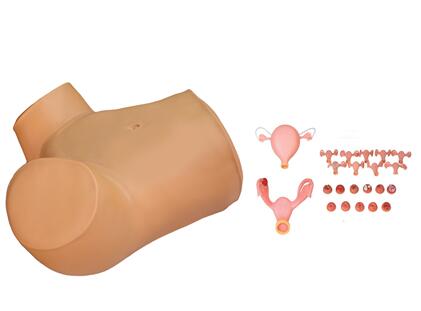
5. Placing and removal of IUD
6. Observe the septal size and position
7. Observe the uterus, ovary, fallopian tube, round
ligament and other structures
8. Normal and abnormal cervix models
1) normal cervix
2) IUD placing and removal from normal cervix
4) cervical laceration
5) cervical polyp
6) cervical inflammation Naboth cyst
7) acute cervicitis
8) cervical adenocarcinoma
9) trichomonad cervicitis
10) cervical verruca acuminata
4) uterus with right salpingitis
5) uterus with right oviduct tubercle